
August 14, 2026
September 2025 – by Tatsuro Misu
Department of Neurology, Tohoku University Hospital, 1-1 Seiryomachi, Aobaku, Sendai 980-8574, Japan
(This article belongs to the Special Issue 25th Anniversary of IJMS: Updates and Advances in Molecular Pathology, Diagnostics, and Therapeutics)
Abstract
Myelin oligodendrocyte glycoprotein (MOG) antibody-associated disease (MOGAD) is characterized by the predominance of optic neuritis, myelitis, acute disseminated encephalomyelitis (ADEM), and cortical encephalitis, and can be diagnosed by the presence of pathogenic immunoglobulin G (IgG) antibodies targeting the extracellular domain of MOG in the serum and cerebrospinal fluid (CSF).
Initially considered a variant of multiple sclerosis (MS) or neuromyelitis optica spectrum disorder (NMOSD), it is now widely recognized as a separate entity, supported by converging evidence from serological, pathological, and clinical studies. Patients with MOGAD often exhibit better recovery from acute attacks; however, their clinical and pathological features vary based on the immunological role of MOG-IgG via antibody- or complement-mediated perivenous demyelinating pathology, in addition to MOG-specific cellular immunity, resulting in heterogeneous demyelinated lesions from vanishing benign forms to tissue necrosis, even though MOGAD is not a mild disease.
The key is the immunological mechanism of devastating lesion coalescence and long-term degenerating mechanisms, which may still accrue, particularly in the relapsing, progressing, and aggressive clinical course of encephalomyelitis. The warning features of the severe clinical forms are: (1) fulminant acute multifocal lesions or multiphasic ADEM transitioning to diffuse (Schilder-type) or tumefactive lesions; (2) cortical or subcortical lesions related to brain atrophy and/or refractory epilepsy (Rasmussen-type); (3) longitudinally extended spinal cord lesions severely affected with residual symptoms.
In addition, it is cautious for patients refractory to acute stage early 1st treatment including intravenous methylprednisolone treatment and apheresis with residual symptoms and relapse activity with immunoglobulin and other 2nd line treatments including B cell depletion therapy. Persistent MOG-IgG high titration, intrathecal production of MOG-IgG, and suggestive markers of higher disease activity, such as cerebrospinal fluid interleukin-6 and complement C5b-9, could be identified as promising markers of higher disease activity, worsening of disability, and poor prognosis, and used to identify signs of escalating treatment strategies. It is promising of currently ongoing investigational antibodies against anti-interleukin-6 receptor and the neonatal Fc receptor.
Moreover, due to possible refractory issues such as the intrathecal production of autoantibody and the involvement of complement in the worsening of the lesion, further developments of other mechanisms of action such as chimeric antigen receptor T-cell (CAR-T) and anti-complement therapies are warranted in the future.
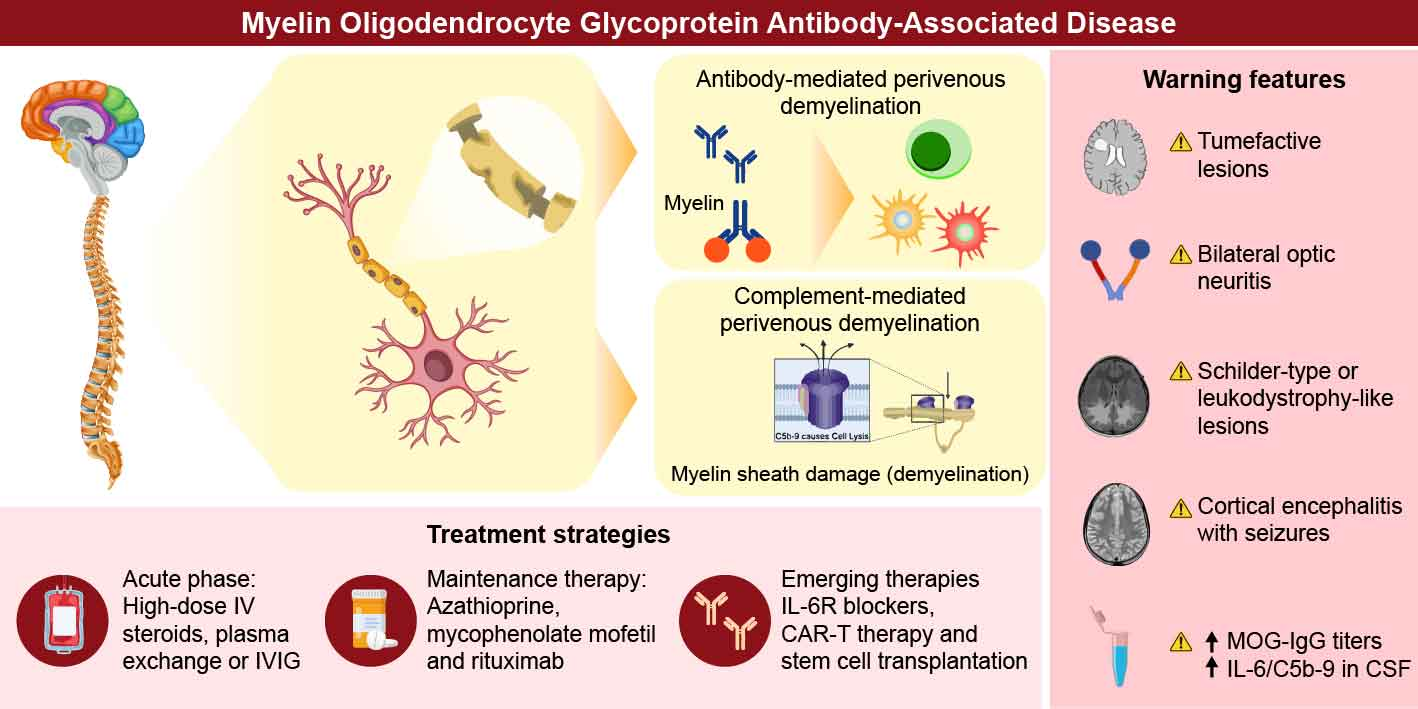
Graphical Abstract
1. Introduction
Myelin oligodendrocyte glycoprotein antibody-associated disease (MOGAD) is a distinct demyelinating disorder of the central nervous system (CNS) characterized by the presence of pathogenic immunoglobulin G (IgG) antibodies targeting the extracellular domain of myelin oligodendrocyte glycoprotein (MOG), a glycoprotein expressed on the surface of oligodendrocytes and the outermost lamellae of CNS myelin sheaths. Initially considered a variant of multiple sclerosis (MS) or neuromyelitis optica spectrum disorder (NMOSD), MOGAD is now widely recognized as a separate nosological entity, supported by converging evidence from serological, pathological, and clinical studies [1,2].
Clinically, MOGAD manifests as diverse phenotypes, including optic neuritis (ON), transverse myelitis (TM), acute disseminated encephalomyelitis (ADEM), and brainstem encephalitis. Notably, disease presentation can vary by age group; children more commonly exhibit ADEM-like symptoms, whereas adults typically present with ON or TM [3,4]. These attacks are often monophasic, but may recur, with some patients experiencing frequent and disabling relapses. Unlike patients with AQP4-IgG+ NMOSD, those with MOGAD often exhibit better recovery from acute attacks. However, long-term disabilities may still occur, particularly in relapsing forms.
Serologically, MOG-IgG is most reliably detected using cell-based assays that use full-length human MOG as the target antigen. These antibodies are predominantly of the IgG1 subtype and thought to be pathogenic via complement- and antibody-dependent cellular cytotoxicity [5,6]. However, immunopathological mechanisms underlying MOGAD remain unclear. Autopsy and biopsy findings reveal perivenous demyelination, axon preservation, and a relative lack of astrocyte loss, contrasting with AQP4-NMOSD [7,8]. These findings, along with distinct cerebrospinal fluid (CSF) profiles and magnetic resonance imaging (MRI) characteristics, reinforce the nosological distinction between MS and NMOSD.
Epidemiological studies have suggested that MOGAD accounts for a significant proportion of acquired demyelinating syndromes, particularly in children with ADEM/encephalitis and adults with AQP4-IgG seronegative NMOSD [9,10]. The annual incidence is approximately 1.6–3.4 per million, although the true prevalence is likely underestimated owing to diagnostic challenges and evolving serological testing standards [5,11]. The MOGAD burden is compounded by its unpredictability; although many patients respond well to steroids and immunotherapies, others experience recurrent or severe attacks that are difficult to control [5,12].
Owing to this clinical heterogeneity, growing attention has been paid to the severe and treatment-refractory subtypes of MOGAD. These forms may be associated with persistent or relapsing disease, poor steroid responsiveness, and atypical imaging patterns. A deeper understanding of immunopathological mechanisms, including the role of intrathecal MOG-IgG synthesis and complement activation, is essential for improving diagnostic precision and tailoring therapy. This review aims to summarize the current knowledge on the pathophysiological basis, imaging characteristics, immunological biomarkers, and therapeutic challenges of refractory and severe MOGAD, to guide future clinical and translational research.
2. Pathological Insights
2.1. Pathological Features of MOGAD
MOGAD is pathologically distinct from MS and aquaporin-4 (AQP4)-IgG+ NMOSD and exhibits characteristic features that reflect its unique immunopathogenesis [13]. Perivenous demyelination and the dominance of CD4+ T-cell infiltration [7,14], along with granulocytes such as neutrophils and eosinophils, often reminiscent of monophasic ADEM, are considered hallmarks of MOGAD in the acute stage [15].
These lesions typically originate around small venules, and complement deposition is relatively rare in MOG antibody+ ADEM cases [7] compared to NMOSD cases; however, some ADEM-like cases resemble type II MS pathology [16]. Unlike the sharply demarcated confluent plaques seen in prototypic MS, MOGAD lesions tend to be ill-defined and patchy, with relative preservation of axons and oligodendrocytes, particularly early lesions [7,8].
This contrasts with the pathology in MS, where CD8+ T cells are predominant and chronic microglial activation occurs in chronically expanding lesion limbs, and in AQP4-NMOSD, which shows marked astrocyte loss and complement-mediated tissue necrosis [17]. These histopathological differences are associated with favorable clinical recovery in patients with MOGAD; clinically relevant T2-lesions in MOGAD resolve more completely and frequently than those in MS and NMOSD [18].
2.2. Smoldering Confluent MS-like Lesions Are Rare in MOGAD
Stay in the loop
Get new articles as they publish
Join our community and we will send trusted education and updates for your condition.